Patellar Luxation in Small Breed Dogs
by Teri Dickinson, DVM
Luxated patellas or "slipped stifles" are a common orthopedic problem
in small dogs. A study of 542 affected individuals revealed that dogs
classified as small (adult weight 9 kg (20 lbs) or less) were twelve
times as likely to be affected as medium, large or giant breed dogs.(1)
In addition, females were 1.5 times as likely to be affected.1
Some researchers have suggested a recessive method of inheritance,(2),(3)
and the higher incidence in females could possibly be related to
X-linked(4) factors or hormonal
influences.
Luxated patellas are a congenital8 (present at birth)
condition. The actual luxation may not be present at birth, but the
structural changes which lead to luxation are present. Most researchers
believe luxated patellas to be heritable (inherited) as well, though the
exact mode of inheritance is not known. The condition is commonly seen
in Italian Greyhounds, although no published data regarding the
incidence in IG's exists at this time. Researchers1 have
suggested that due to the high risk factor in toy breeds, breeding
trials or retrospective pedigree analyses should be undertaken by
national breed clubs to answer some of these questions.
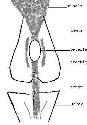 The
stifle is a complicated joint(5) which is
the anatomical equivalent of the human knee. The three major components
involved in luxating patellas are the femur (thigh bone), patella (knee
cap), and tibia (calf or second thigh). See drawing A. In a normal
stifle, the femur and tibia are lined up so that the patella rests in a
groove (trochlea) on the femur, and its attachment (the patellar tendon)
is on the tibia directly below the trochlea. The
stifle is a complicated joint(5) which is
the anatomical equivalent of the human knee. The three major components
involved in luxating patellas are the femur (thigh bone), patella (knee
cap), and tibia (calf or second thigh). See drawing A. In a normal
stifle, the femur and tibia are lined up so that the patella rests in a
groove (trochlea) on the femur, and its attachment (the patellar tendon)
is on the tibia directly below the trochlea.
The function of the patella is to protect the large tendon of the
quadriceps (thigh) muscle as it rides over the front of the femur while
the quadriceps is used to extend (straighten) the stifle joint. Placing
your hand on your patella (knee cap) while flexing and extending your
stifle (knee) will allow you to feel the normal movement of the patella
as it glides up and down in the trochlea.
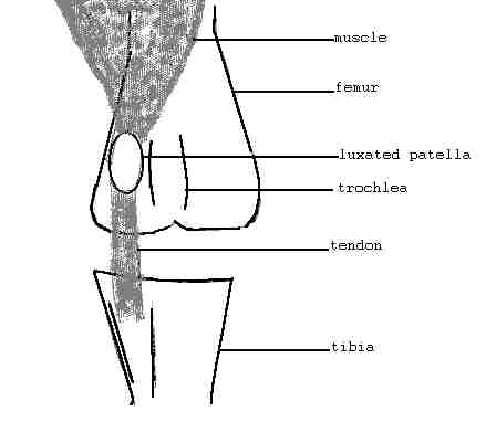 Luxation
(dislocation) of the patella occurs when these structures are not in
proper alignment.(6) Luxation in toy
breeds most frequently occurs medially (to the inside of the leg). See
drawing B. The tibia is rotated medially (inward) which allows the
patella to luxate (slip out of its groove) and ride on the inner surface
of the femur. Luxation
(dislocation) of the patella occurs when these structures are not in
proper alignment.(6) Luxation in toy
breeds most frequently occurs medially (to the inside of the leg). See
drawing B. The tibia is rotated medially (inward) which allows the
patella to luxate (slip out of its groove) and ride on the inner surface
of the femur.
While the patella is luxated, the quadriceps is unable to properly
extend the stifle, resulting in an abnormal gait or lameness. In
addition, the smooth surface of the patella is damaged by contact with
the femur, rather than the smooth articular (joint) cartilage present in
the trochlea. With time this rubbing will result in degenerative joint
disease (arthritis). Furthermore, while the patella is luxated, the
quadriceps puts a rotational force on the tibia, which over time will
increase the rotation of the tibia, thereby increasing the severity of
the problem. The additional strain caused by the malformation of the
bones may also lead to later ligament ruptures. Many individuals are
affected bilaterally (both legs).
Signs of luxation may appear as early as weaning or may go undetected
until later in life. Signs include intermittent rear leg lameness, often
shifting from one leg to the other, and an inability to fully extend the
stifle. The leg may carried for variable periods of time. Early in the
course of the disease, or in mildly affected animals, a hopping or
skipping action occurs. This is due to the patella luxating while the
dog is moving and by giving an extra hop or skip the dog extends its
stifle and is often able to replace the patella until the next luxation,
when the cycle repeats.
Several grades of luxation have been defined(7),5.
In simple terms they are:
- Grade I. Patella can be luxated manually (by the examiner) but
returns to the trochlea when released. Occasional luxation occurs
causing the animal to temporarily carry the limb. Tibial rotation is
minimal
- Grade II. Patella can be easily luxated manually and remains
luxated until replaced. Luxation occurs frequently for longer
periods of time, causing the leg to be carried or used without full
extension. Tibial rotation is present.
- Grade III. The patella is permanently luxated, but can be
replaced manually. The dog often uses the leg, but without full
extension. Tibial rotation is marked.
- Grade IV. The patella cannot be replaced manually, and the leg
is carried or used in a crouching position. Extension of the stifle
is virtually impossible. Tibial rotation is quite severe, resulting
in a "bow legged" appearance.
While no data has been published, personal observation reveals most
affected IG's appear to have Grade I or II luxations. I have also
encountered puppies born with no trochlea and severe tibial rotation
causing permanent luxation from birth (Grade IV), and adult dogs so
severely affected they were non-weight bearing in both hind legs and
merely dragged their rear legs along in a frog-like position (Grade IV).
Diagnosis is relatively simple for a veterinarian familiar with
orthopedics. It involves palpation of the joint and manual luxation of
the patella. X-rays may also be used to determine the degree of
rotation. Motivated owners may be trained by veterinarians to palpate
the stifles, but care must be exercised in order to avoid injuring the
joint, or making an incorrect diagnosis.
Diagnosis in severe cases may be possible at weaning, but in most cases
the joints should be tight enough at 4 to 6 months(8)
to allow reliable palpation. Screening of puppies at this age will help
prevent large expenditures training and showing dogs which later prove
unsound. Screening of breeding stock and culling of affected individuals
should, over time, reduce the incidence of the condition.
Treatment involves surgical correction of the deformities. Many
techniques are available depending on the severity of the condition.(9)
Satisfactory results are usually obtained if the joint degeneration has
not progressed too far. Once the condition is repaired, most affected
individuals make satisfactory pets.
Bibliography
1. Priester WA: Sex, Size and Breed as Risk
Factors in Canine Patellar Luxation. J Am Vet Med Assoc. 160:740, 1972.
2. Hutt FB: Genetic Defects of Bones and Joints in
Domestic Animals. Cornell Vet. 58:104, 1968.
3. Kodituwakku GE: Luxation of the Patella in the
Dog. Vet. Rec. 74:1499, 1962.
4. present on the X chromosome, of which females
have two, XX and males one, XY
5. Miller, ME: Anatomy of the Dog. WB Saunders
Co., Philadelphia, PA 1964.
6. Putnam RW: Patellar Luxation in the Dog. M.Sc.
Thesis. Presented to the faculty of graduate studies, University of
Guelph, Ontario, Canada, January 1968.
7. Singleton WB: The Surgical Correction of Stifle
Deformities in the Dog. J Small An Pract 10:59, 1969.
8. Archibald J: Canine Surgery. American
Veterinary Publications, Santa Barbara, CA, 1974.
9. Brinker WO: Handbook of Small Animal
Orthopedics & Fracture Treatment. WB Saunders Co., Philadelphia, PA,
1990.
|